61 High Street Ware SG12 9AD | Click here for map

Optical coherence tomography, also called OCT, is a method for imaging the various layers of the retina, the optic nerve head, and the anterior segment of the eye. More advanced than retinal photography (which can only view the surface of the retina) the OCT enables us to view the individual layers. Similar to ultrasound, individual layers of tissue are scanned without touching the eye and displayed as a cross-section. A completely safe light source is used. A typical example of an OCT image of the retina is shown below. The examination lasts a few seconds, does not require the use of drugs or drops, and is totally painless.
 OCT supports your optometrist in diagnosing a disease and monitoring change over time. The OCT is particularly useful for issues concerning fluid retention and swelling in the retina, which can occur, for example, with age-related macular degeneration (AMD) or diabetes. But very early signs of a pathological change can also be detected.
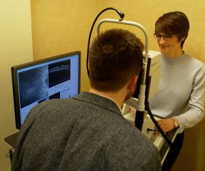 The OCT scan is so detailed that it can pick up the very earliest signs of glaucoma – damage to the microscopic nerve fibres at the back of your eye. This makes it possible to detect the very early signs of glaucoma before it affects your sight, and indeed before it is detectable using more traditional techniques. This makes OCT the definitive screening tool currently available for this condition.
The OCT also provides us with highly useful information about other parts of your eye such as small retinal holes, tears or cysts that may be present.





Copyright © 1947-2026
Aves Ware LLP T/A Aves Optometrists • Registered No: OC454839 • Registered Address: Aves, 61 High Street, Ware, Hertfordshire, SG12 9AD
All rights reserved.

